This post is an answer to the Case – Polyarthralgia and Painless Subcutaneous Nodules
A bone marrow biopsy was performed, which was normocellular. An x-ray revealed severe erosion and deformities of the hand bones and punched-out bony radiolucencies associated with the subcutaneous nodular lesions (figure B).
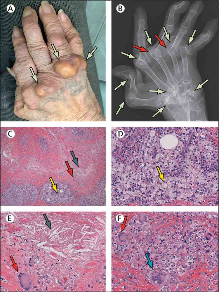
A skin biopsy specimen of the subcutaneous mass showed areas of cholesterol clefts and necrobiosis surrounded by foamy histiocytes, lymphocytes, Touton giant cells, and foreign body-type multinucleated giant cells (figure C–F). These findings confirmed a diagnosis of pseudoxanthomatous rheumatoid nodule.
Pseudoxanthomatous rheumatoid nodules are an unusual form of rheumatoid nodule resembling tendon xanthomas, are often mistakenly diagnosed as xanthomatosis, and have been previously regarded by investigators as subcutaneous xanthogranuloma, observed in the juxta-articular sites of the hand, wrist, elbow, knee, foot, and acromioclavicular joints.
Pseudoxanthomatous rheumatoid nodules are distinguished from xanthomatosis by x-ray findings of demonstrable punched-out bony radiolucencies, which are not present in xanthomatosis. Cholesterol clefts surrounded by granulomatous reactions can also be observed in rheumatoid synovial fluid and bursitis, sometimes presenting as migrating chylous cysts.
Pseudoxanthomatous rheumatoid nodule and xanthomatosis are easily misdiagnosed because their histopathological findings are similar. However, xanthomatosis is often associated with hyperlipidaemia, cryoglobulinemia, hypocomplementemia, and monoclonal paraproteins, whereas pseudoxanthomatous rheumatoid nodule is not accompanied by these diseases and is found in the juxta-articular sites with punched-out bony radiolucencies.
After initiating treatment with 6 mg oral methotrexate per week, the concentrations of CRP reduced to 0·63 mg/dL, polyarthralgia improved, and the pseudoxanthomatous rheumatoid nodules shrank.