This post is an answer to the ECG Case 237
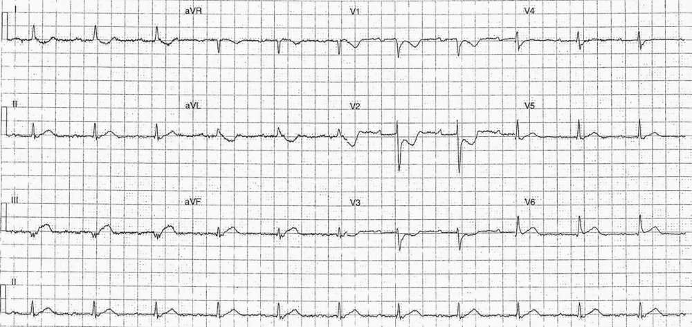
- Rate: 66 bpm
- Rhythm:
- Regular
- Sinus rhythm
- Baseline artifact makes P waves difficult to see but best seen in leads V1-3
- Axis: Normal
- Intervals:
- PR – Prolonged (280ms)
- QRS – Normal (80-100ms)
- QT – 380ms (QTc Bazette 400 ms)
- Segments:
- ST Elevation in leads III, aVF (<1mm)
- Flat ST depression in leads V1-3
- Additional:
- T wave inversion in leads I, aVL, aVR, V1-3
- Prominent T waves in leads III, aVF, V6
- Prominent R wave in lead V2
Interpretation
Infero-postero-lateral MI
What happened next ?
The patient was transferred for urgent PCI and a lesion was stented. Unfortunately the hospital discharge summary doesn’t state where the culprit lesion was.
SIMILAR CASES:


