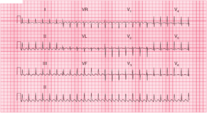
ECG Interpretation
- Sinus rhythm, rate 64/min
- Normal axis
- Q (QS) waves in leads V1–V4
- ST segment Elevation in leads V1–V4, and subtle ST elevation in aVL
- Reciprocal ST Depression in inferior leads (II,III,aVF) and in leads V5 and V6
- Inverted T waves in leads I, aVL, V2–V6
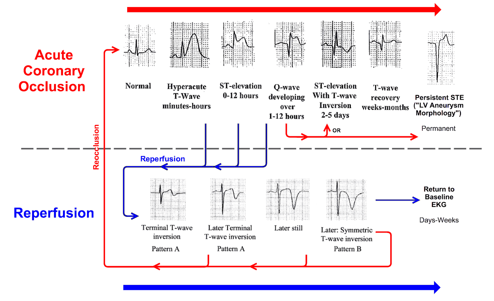
- Terminal T wave inversion in leads V2-V4 indicating spontaneous reperfusion
Clinical Interpretation
This is a classic acute ST segment elevation anterior myocardial infarction (STEMI) with spontaneous reperfusion indicated by the Terminal T wave inversion in leads V2-V4.
What to do next ?
More than 18 h have elapsed since the onset of pain, so this patient is outside the conventional limit for thrombolysis or percutaneous coronary intervention (PCI).
Nevertheless, if he is still in pain and still looks unwell, PCI or thrombolytic treatment should be given unless there are good reasons not to do so. In any case, he should be given pain relief and aspirin, and must be admitted to hospital for observation.