This post is an interpretation of the ECG Case 195
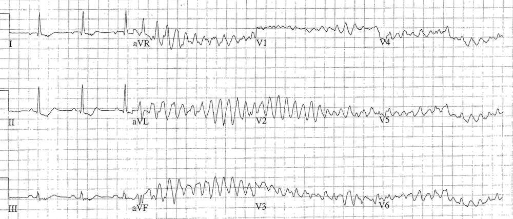
Interpretation of the Initial 3 Complexes
- Rate: ~65-68 / min
- Rhythm: Regular
- Axis: Normal
- Intervals:
- PR – Normal (~160ms)
- QRS – Normal (100ms)
- QT – 400ms (QTc Bazette ~420-430 ms)
- Segments:
- ST Depression in leads I, II, III
- Additional: P Wave Inversion in Lead I
- Interpretation: Ectopic Atrial Rhythm with ischemic features
Interpretation of Subsequent ECG
- Ventricular Ectopic with ‘R-onT’ phenomenon
- Polymorphic VT then VF
Interpretation: Acute myocardial ischaemia / infarction causing polymorphic VT / VF
What happened next ?
- CPR
- Received 4 x 200J shocks
- 150mg iv amiodarone
- 100 mg iv lignocaine
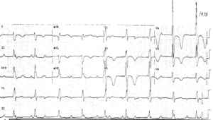
Subsequent ROSC was achieved after less than 10 minutes. Post ROSC ECG showed antero-lateral ST elevation.
The patient underwent inter-hospital transfer for PCI. PCI revealed a proximal LAD lesion with 90% occlusion, which was stented.
Echo showed:
- Normal LV size with anterior, septal and apical akinesis and overall moderate systolic impairment
- Probable LV apical thrombus
- Normal right ventricular size and apical akinesis and overall mild systolic impairment.
The patient was subsequently discharged on warfarin, anti-platelet therapy, ACE inhibitor, beta-blocker, and a statin.
SIMILAR CASES:


![Read more about the article Atrial Flutter: ECG Interpretation [With Examples]](https://manualofmedicine.com/wp-content/uploads/2022/01/Atrial-Flutter-with-4-1-AV-Block-300x135.png)
