This post is an answer to the ECG Case 290
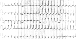
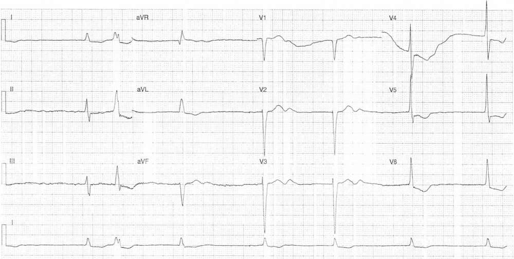
- Rate: 42 bpm
- Rhythm: No p waves visible
- Axis: Normal (14 deg)
- Intervals:
- QRS – Prolonged (110-120ms)
- Segments:
- ST Depression in leads I, II, aVL, V4-6
- Additional:
- Prominent U waves in leads V1-3
- Single ectopic beat
- LBBB Morphology
Intepretation
- Slow atrial fibrillation with ventricular ectopic
- Differentials include:
- Sinus node dysfunction
- Drug toxicity – digoxin, CCB, beta-blocker
- Hypothermia
- Hypothyroid
- Ischemia
- Differentials include:
- Lateral ST depression
- Differentials include
- Digitalis effect
- Electrolyte abnormality
- Ischemia
- Differentials include
What happened ?
The patient was admitted under the cardiology team. Following liaison with his GP it was discovered the patient was on digoxin and apixaban. Serum digoxin levels were normal as were potassium levels and despite several days of observation the patient had persistent bradycardia with intermittent junctional escape rhythms. Following an echo which showed an EF of >50% the patient underwent an uneventful PPM insertion.
SIMILAR CASES: