This post is an answer to the ECG Case 320
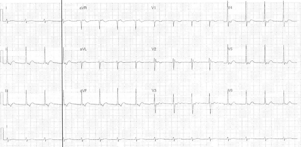
- Rate 96 bpm
- Regular narrow complex
- Partial RBBB pattern rSr’ in lead V2
- Right axis deviation
- Retrograde P waves
- Inverted in leads II, III, aVF, V4-6
- Nil ST / T wave abnormality accounting for disruption due to retrograde P waves
Impression:
- Accelerated junctional rhythm
Accelerated junctional rhythms occur when the rate of the AV nodal pacemaker cells exceed that of the sinus node. This can occur in settings of decreased sinus node activity, excessive vagal tone or increased AV nodal automaticity. They are not always pathological and can occur in the setting of sleep, young healthy individuals, and times of high vagal tone. They can also been seen in the setting of digoxin toxicity, ischaemia, myocarditis, post-cardiac surgery and due to drug effects.
What happened next ?
The patient had negative serial troponins and a negative D-dimer. She was admitted under cardiology and had a normal echocardiogram. There were no further episodes of chest pain during telemetry monitoring.
SIMILAR CASE: Junctional Rhythm