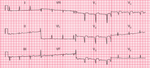
This post is an answer to the ECG Case 344
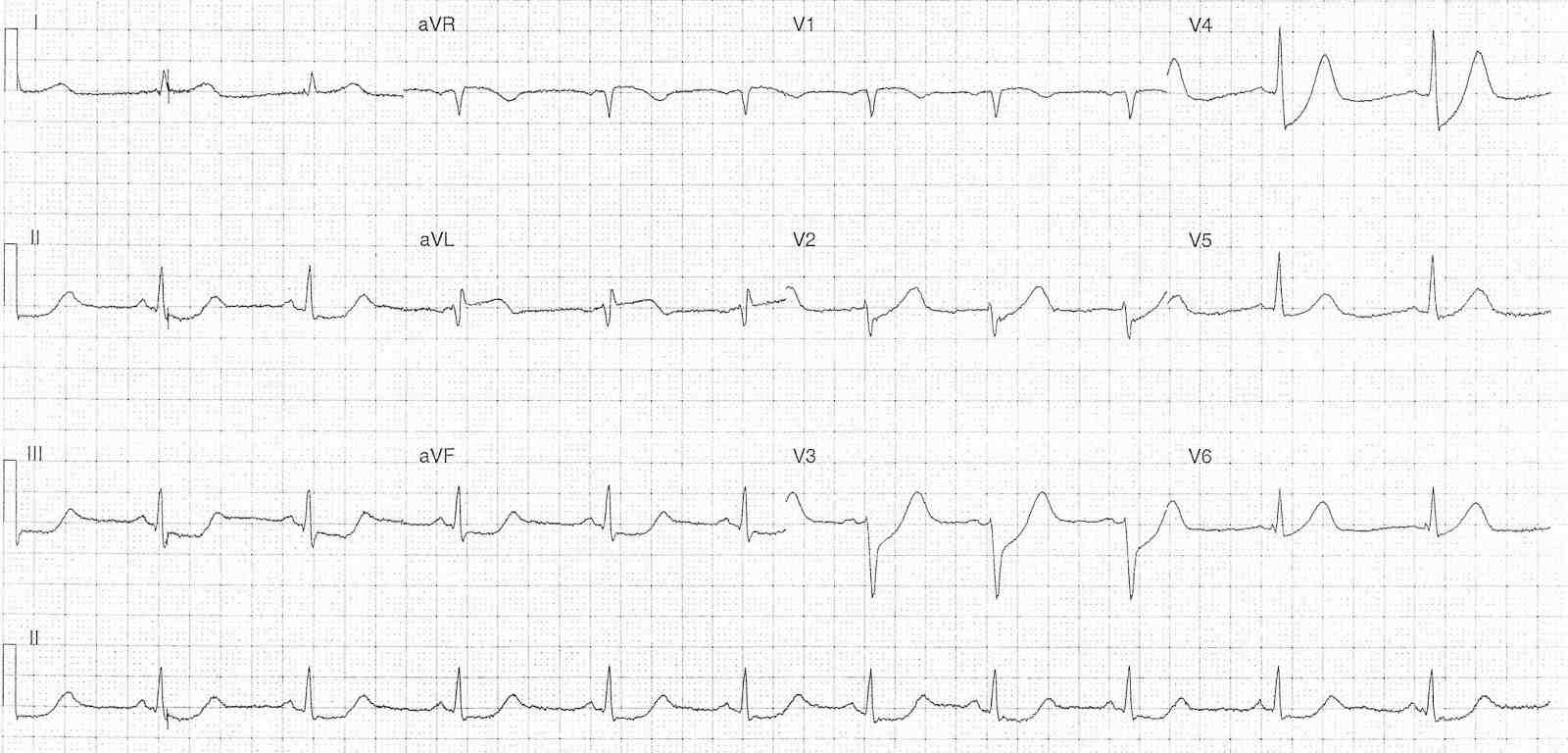
- Rate: 60 bpm
- Rhythm: regular, sinus rhythm
- Axis: Normal
- Intervals:
- PR – Normal
- QRS – Normal
- Additional:
- ST Elevation in leads aVR and aVL (<1mm)
- Upsloping ST depression in leads V2-6
- Maximal in leads V3-4
- Flat ST depression in the inferior leads
- Prominent T waves in leads V2-4
Interpretation
Features consistent with a De Winter’s pattern, suggesting potential LAD pathology.
Given the patients age and medical history broader differentials for shock and ischaemic ECG features would be:
- PE
- Sepsis
- Endocrine – Sheehan’s Syndrome
What happened next ?
The patient was initially treated with iv fluid, analgesia and broad spectrum iv antibiotics (as initial broad DDx included sepsis). Following review of the initial ECG urgent cardiology input was obtained and an emergent CTPA was performed to exclude PE as a potential cause.
On return from a negative CTPA the patient complained of further chest pain and was transferred for urgent angiography which showed a 90% mid LAD lesion treated with bioresorbable stent. Subsequent CTPA revealed a coronary artery dissection as the cause for the acute LAD lesion.
Coronary Artery Dissection
This is a rare phenomena occurring in ~4% of all acute MI’s. Approximately 90% of cases occur in females and it accounts for 1/4 of MI’s in the under 50-year-old age group. Recurrence rate is between 13-18%. There are multiple risk factors including:
- Fibromuscular dysplasia
- Pregnancy
- Connective tissue disorders
- Systemic inflammatory conditions
- Intense exercise
- Intense emotional stress
- Labour and delivery
- Valsalva-like events
- Sympathomimetic use
SIMIALAR CASES: