This post is an answer to the Case – Chest Pressure After Eating
Findings
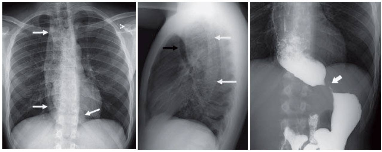
- Chest radiograph shows a tubular soft tissue density mass (white arrows) with mottled air extending through the middle mediastinum from the thoracic inlet to the diaphragm. The trachea is deviated anteriorly (black arrow).
- Upper gastrointestinal study confirms the marked dilatation of a debris-filled esophagus with beak-like tapering (thick white arrow) at the gastroesophageal junction. Intermittent emptying of the esophagus into the stomach was noted at the time of the exam, as well as tertiary contractions.
Differential Diagnosis
The chest radiograph finding can only be explained by esophageal dilatation, the differential for which consists of achalasia, a distal esophageal stricture, or an obstructing esophageal or gastric mass. The demonstration of intermittent opening on fluoroscopy and the beak-like tapering would support the diagnosis of achalasia. Occasionally, scleroderma may result in a patulous esophagus, which does not usually reach such a large size.
Teaching Points
- A primary esophageal disorder of unclear etiology, achalasia results from destruction of the myenteric plexus, which impairs normal esophageal peristalsis and lower esophageal sphincter function. This results in progressive retention of food products and fluid within the esophagus.
- On barium swallow, there is absence of primary and secondary esophageal peristalsis and smooth tapering of the esophagus at the gastroesophageal junction with intermittent relaxation. Esophageal manometry is considered the gold standard for diagnosis.
- Rarely, Chagas disease can result in a radiographic appearance similar to achalasia.
- Achalasia is associated with a higher rate of nontuberculous mycobacterial infection.
Management
A range of treatment options exist, including balloon dilatation, oral medications, botulinum toxin injection, and surgical myotomy. Referral to a gastroenterologist or surgeon is often indicated.
Further Reading
- Franquet T, Erasmus JJ, Giménez A, Rossi S, Prats R. The retrotracheal space: normal anatomic and pathologic appearances. Radiographics. 2002 Oct;22 Spec No:S231-46.
- Pohl D, Tutuian R. Achalasia: an overview of diagnosis and treatment. J Gastrointestin Liver Dis. 2007 Sep;16(3):297-303. PMID: 17925926.