This article is an answer to the Case – Woman with Dermatomyositis and Subcutaneous Nodules on the Arms and Legs
At the time of the initial diagnosis 20 years earlier, manifestations of dermatomyositis had included an inflammatory myopathy, Gottron’s papules, and both a heliotrope and photosensitive rash.
Treatment with methotrexate and prednisone had resulted in remission of the myopathy, but she continued to have active inflammatory skin disease, and during the past 10 years she had progression of hard, mobile, painless subcutaneous nodules.
Serum levels of ionized calcium, phosphorus, albumin, 25-hydroxyvitamin D, and intact parathyroid hormone were normal.
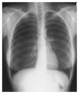
A radiograph of the left shoulder showed extensive soft-tissue calcification and an old fracture of the surgical neck of the humerus, which was attributed to glucocorticoid-induced osteoporosis.
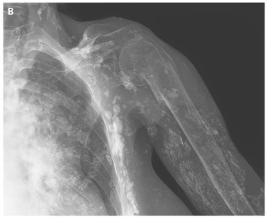
The clinical presentation was consistent with a diagnosis of dermatomyositis-associated calcinosis. In addition to its association with dermatomyositis, soft-tissue calcinosis can be seen in other connective-tissue disorders, such as systemic sclerosis (scleroderma). The calcifications progressed during treatment with infliximab.