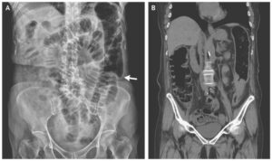
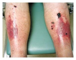
This article is an answer to the Case – Generalized Pustular Eruptions
Laboratory studies indicated neutrophilia and mild eosinophilia (with 55% neutrophils and 8% eosinophils). The white-cell count was 6500 per cubic millimeter.
Histopathological examination of the lesions revealed pseudoepitheliomatous hyperplasia, full-thickness epidermal necrosis, and a diffuse neutrophilic dermal infiltrate. The results of microbiologic studies were negative. On the basis of these findings, a diagnosis of iododerma was made.
Iododerma is a hypersensitivity reaction to iodine. Although its pathogenesis is not understood, delayed iodine clearance and the induction of neutrophil degranulation have been proposed as mechanisms.
Intravenous injection of iodinated contrast material is among the most common causes of iododerma; however, iododerma has also been reported after the oral or topical administration of iodine.
Treatment with thalidomide was initiated, and the skin lesions completely resolved within 4 weeks. After careful evaluation, the cause of the hematuria was not identified. The hematuria resolved spontaneously within 3 days without treatment. The patient’s kidney function remained normal.


