This post is an answer to the Case – 35-year-old with Chest Pain and Shortness of Breath
Findings
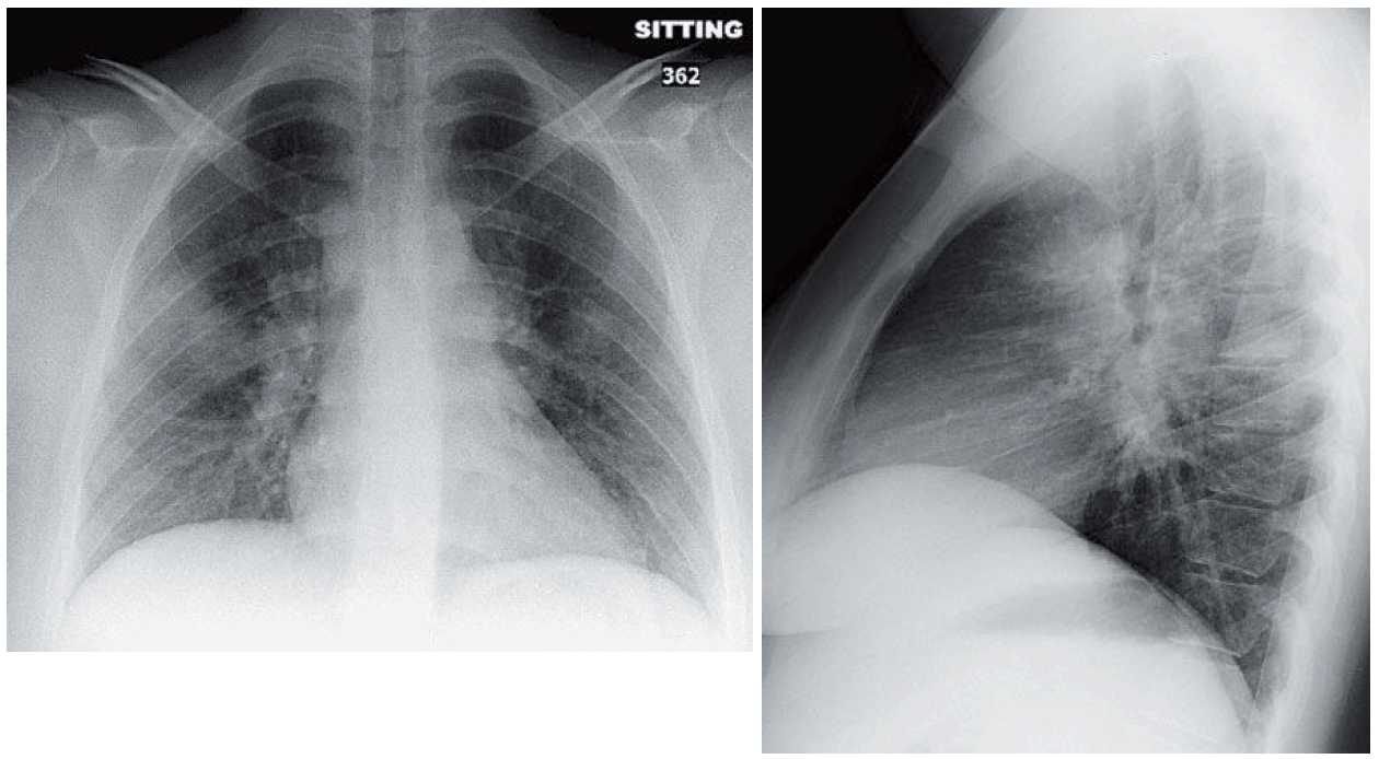
- The frontal radiograph demonstrates bilateral lobulated hila in keeping with hilar lymphadenopathy. Right paratracheal lymphadenopathy and aorticopulmonary lymphadenopathy are also seen.
- The lateral radiograph demonstrates a circular opacity surrounding the mainstem bronchi (doughnut sign). This ring is in keeping with middle mediastinal lymphadenopathy.
Differential Diagnosis
Mediastinal lymphadenopathy can be seen in the setting of granulomatous infection, granulomatous inflammation (sarcoid), or neoplasm (lymphoma or metastases). The homogenous attenuation, lack of vascular occlusion, symmetry in the mediastinum, parenchymal opacities, and young age would favor sarcoidosis.
Teaching Points
- On a chest radiograph, the right paratracheal stripe should be <4 mm. When larger, a mediastinal mass (usually lymphadenopathy) should be suspected.
- The aorticopulmonary window (the left mediastinal border between the aorta and main pulmonary artery) should be concave. When it is convex, a mediastinal mass (usually lymphadenopathy) should be suspected.
- Distinguishing enlarged pulmonary arteries from hilar lymphadenopathy can be challenging. The visualization of an inferior border is a useful finding in lymphadenopathy; it is not seen with enlarged vessels.
- Lymphadenopathy may be easily missed on the frontal examination, but using the lateral examination to look for the doughnut sign can be helpful.
- CT is useful in determining the location and pattern of the lymphadenopathy and lung findings that will direct the differential diagnosis. Calcification, enhancement, and low attenuation are features that can help refine the differential diagnosis on CT.
- Sarcoidosis tends to involve the right paratracheal and bilateral lymph nodes. This is sometimes referred to as a one-two-three pattern.
- Lymphadenopathy in sarcoidosis tends to be homogeneous and soft tissue in attenuation without compression of adjacent vasculature or bronchi.
Management
Further evaluation in this situation would begin with transbronchial biopsy. Right paratracheal nodes can be sampled by conventional mediastinoscopy. Increasingly, endoscopic bronchoscopic ultrasound (EBUS) is being used for hilar node sampling.
SIMILAR CASE: Patient with Hypercalcemia and Suppressed Parathyroid Hormone Level
Further Reading
Sharma A, Fidias P, Hayman LA, et al. Patterns of lymphadenopathy in thoracic malignancies. Radiographics. 2004 Mar-Apr; 24 (2):419-434.