This article is an answer to the Case – Patient with Postprandial Dyspnea, Epigastric Pain and Substernal Fullness
A stool specimen was negative for occult blood. The results of laboratory tests were as follows: hemoglobin level, 12.8 g per deciliter; hematocrit, 37 percent; mean corpuscular volume, 86 μm3; and white-cell count, 7400 per cubic millimeter.
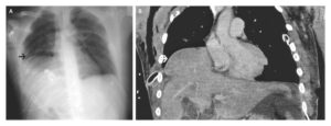
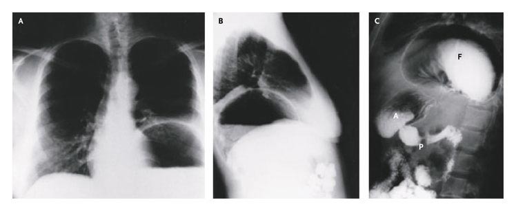
Posteroanterior and lateral chest radiography revealed a large intrathoracic gastric bubble (Panels A and B), and a barium-contrast study of the upper gastrointestinal tract confirmed the presence of a large paraesophageal hernia (Panel C). A denotes gastric antrum, F gastric fundus, and P pylorus.
On endoscopic examination, no hemorrhage, mucosal damage, or esophagitis was found. An exploratory laparotomy revealed a large paraesophageal hernia (i.e., type II hiatal hernia), without evidence of gastric strangulation.
The crura were repaired, and a Nissen fundoplication with anterior gastropexy was performed. The patient recovered from surgery uneventfully.