
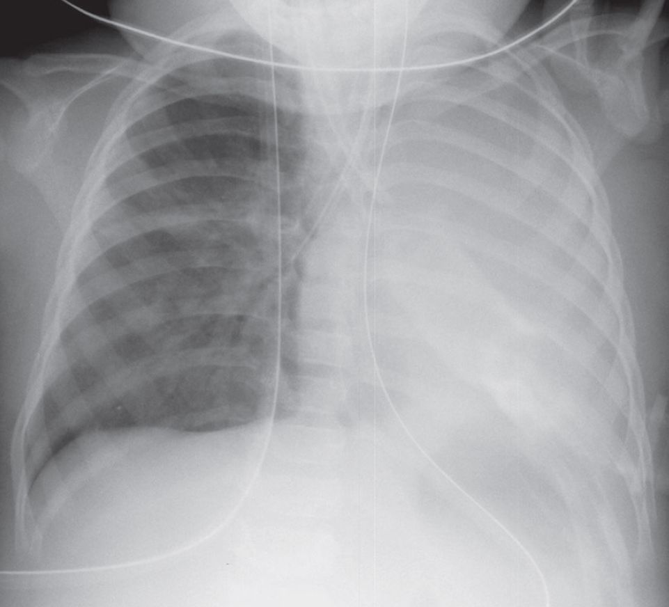
AP chest radiograph demonstrates an opaque left thorax with marked overinflation of the right lung and ipsilateral mediastinal shift. The endotracheal tube is malpositioned, with the tip in the right mainstem bronchus.
Diagnosis
Total Atelectasis Left Lung; Inadvertent Right Mainstem Bronchial Intubation
Differential Diagnosis
- Post-Pneumonectomy
- Massive Hydrothorax
- Extensive Pneumonia
Clinical Findings in Total Atelectasis
Complete atelectasis of a lung is associated with diminished or absent breath sounds in the affected thorax. Patients may experience tachypnea, tachycardia, and an abrupt drop in oxygen saturations.
Imaging Findings
- Opacification of affected thorax
- Ipsilateral mediastinal shift
- Ipsilateral diaphragmatic elevation
- Compensatory overinflation of contralateral lung
Management
- Relieve mainstem bronchial obstruction
- Correction of malpositioned endotracheal tube
Prognosis
- Good if recognized early and promptly treated
- Delayed recognition and treatment may be complicated by barotrauma (e.g., pulmonary interstitial emphysema, pneumomediastinum, spontaneous pneumothorax
Pearls
- Entire lung atelectasis is usually secondary to complete obstruction of a main bronchus.
- Malpositioned ET tube should be excluded in critical care patients with isolated upper lobe or complete lung atelectasis; atelectasis occurs rapidly in such settings.
- Understanding direct and indirect signs of volume loss allows the radiologist to exclude massive unilateral effusion or diffuse unilateral pneumonia.