
This post is an answer to the Case – Growing Mass on the Big Toe
After 8 years of levothyroxine therapy, a male patient who underwent thyroidectomy at the age of 47 years for Graves’ disease developed a 3 cm mass on his left hallux. The lesion was removed and diagnosed as localised myxoedema.
Two years later, at the age of 57 years, the patient presented with a local recurrence. On physical examination, he had orbitopathy and no sign of pretibial myxoedema; the recurrence presented as a soft, mobile lump of the dorsal aspect of the hallux (figure). MRI showed a suprafascial mass with hyperintense T2-weighted signal and poorly defined margins (figure).
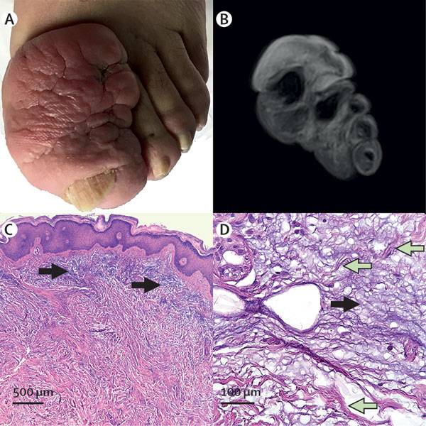
(B) MRI of the recurrent mass showing hyperintense T2-weighted signal and poorly defined margins.
Haematoxylin and eosin staining of the original mass showing basophilic material (black arrows) expanding the reticular dermis and dissecting collagen fibres (green arrows; C, 40x magnification; and D, 200x magnification).
To confirm the dermopathy and to exclude a soft tissue neoplasm, tissue slides of the previous surgery were reviewed. Haematoxylin and eosin stain showed a diffuse widening of the reticular dermis by a faintly basophilic acellular material, associated with isolated lymphocytes, that was dissecting collagen fibres and diffusing in perivascular and periadnexal spaces (figure).
These microscopic features were consistent with an extracellular mucopolysaccharides deposit (ie, myxoedema). The original diagnosis was thus confirmed, the present lesion considered a recurrence, and the patient referred for conservative management.
SIMILAR CASES: