This post is an answer to the Case – Patient with Acute Respiratory Failure and Sepsis
Findings
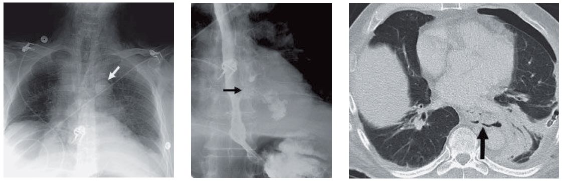
- The chest radiograph demonstrates pneumomediastinum (white arrow in first image) with retrocardiac consolidation and lack of distinction of the descending aorta.
- The esophogram demonstrates extravasation of contrast in the lower third of the esophagus, consistent with a leak (black arrow in second image).
- Subsequent CT confirms perforation of the esophagus. Discontinuity of the wall of the esophagus with pneumomediastinum and left lower lobe pneumonia are seen. Chest tubes were placed because of suspicion for empyema.
Differential Diagnosis
Based on the differential diagnosis of pneumomediastinum in the absence of trauma, the differential diagnosis would include esophageal perforation, tracheal injury, retroperitoneal gas (from diverticular disease), and spontaneous pneumomediastinum. The left lower lobe pneumonia and small effusion would raise the concern for esophageal perforation, which was confirmed on fluoroscopy.
Teaching Points
- Esophageal perforation may occur in a variety of iatrogenic settings: therapeutic endoscopic procedures such as stricture dilation, stent placement, or thermal injury in left atrial radiofrequency ablation.
- Esophageal rupture may occur spontaneously, as in Boerhaave syndrome, in which incomplete cricopharyngeal relaxation during vomiting results in abruptly increased intraluminal pressure sufficient to rupture the esophagus.
- The distal left posterior wall is the most common site of spontaneous rupture, which classically results in pneumomediastinum and left pleural effusion.
- Other possible causes of esophageal perforation include foreign body impaction, caustic and infectious esophagitis, Barrett syndrome, and esophageal cancer.
- A dreaded complication of esophageal perforation is the development of mediastinitis, which can be lethal, but can also result in discitis and fistula formation, most notably with the aorta.
- Although spontaneous pneumomediastinum may present with chest discomfort, this condition should not present with consolidation or effusions.
Management
The optimal treatment of esophageal perforation depends on a host of considerations. Treatment methods range from nonsurgical management to esophagectomy or surgical exclusion and diversion; however, with an early diagnosis of uncontained perforation, surgery remains the mainstay of therapy.
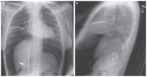
SIMILAR CASE: Severe Pain and Edema of the Neck and Chest
Further Reading
- Fadoo F, Ruiz DE, Dawn SK, Webb WR, Gotway MB. Helical CT esophagography for the evaluation of suspected esophageal perforation or rupture. AJR Am J Roentgenol. 2004 May;182(5):1177-9. doi: 10.2214/ajr.182.5.1821177. PMID: 15100114.
- Young CA, Menias CO, Bhalla S, Prasad SR. CT features of esophageal emergencies. Radiographics. 2008 Oct;28(6):1541-53. doi: 10.1148/rg.286085520. PMID: 18936020.