This post is an answer to the ECG Case 348
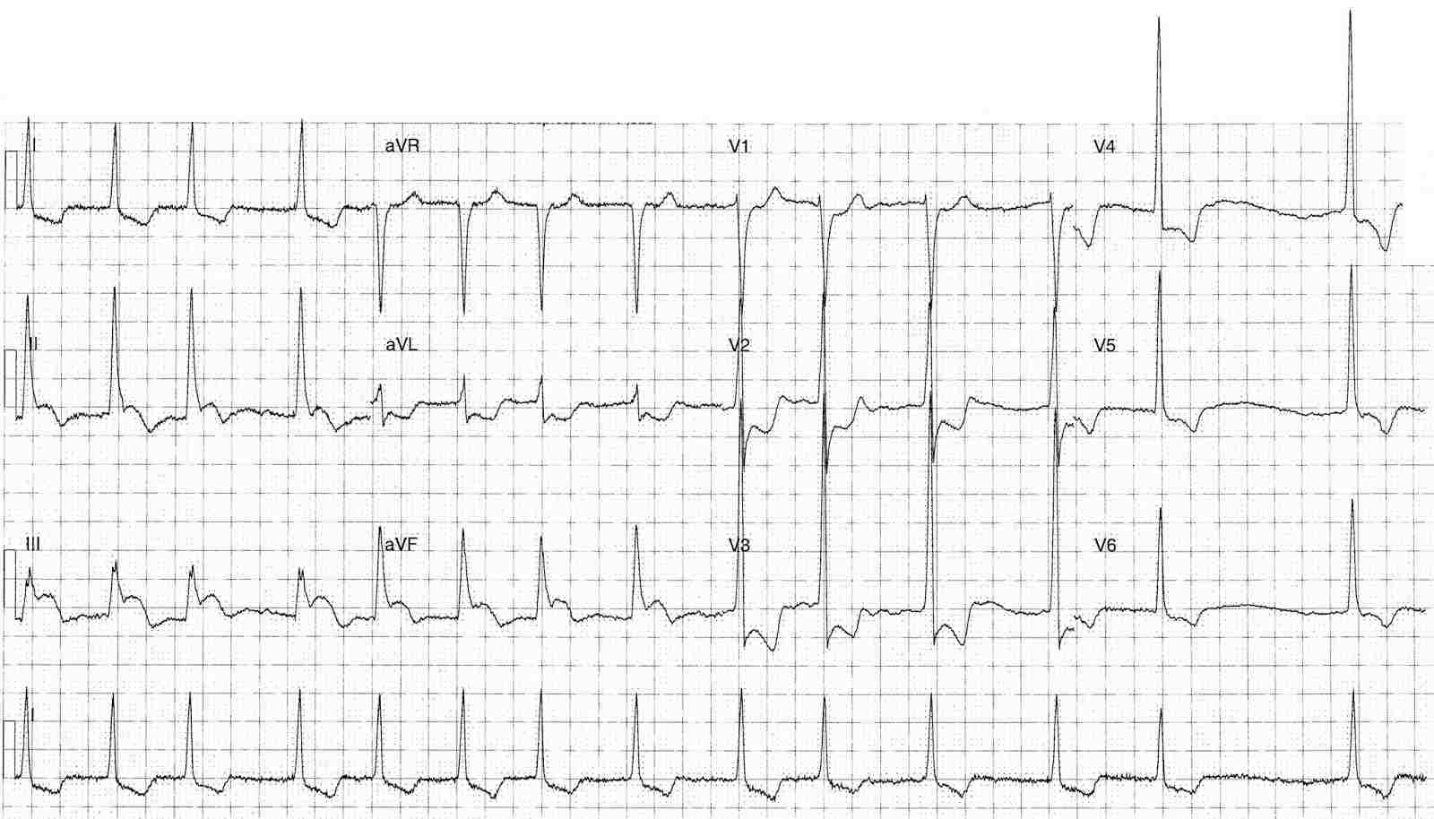
- Rate: Mean ventricular rate 84 bpm
- Rhythm:
- Irregular
- Absent p waves
- Axis: Normal
- Intervals: QRS – Normal (100ms)
- Segments:
- ST Elevation in leads II (1-2mm) III (3mm) aVF (2mm)
- ST Depression in leads I, aVL, V2-6
- Additional:
- T wave inversion in leads I, V2-6
- Biphasic T wave in leads aVL, V2
- R/S ratio V2 >1
- Voltage criteria for LVH
Interpretation
- Infero-posterior OMI
- Atrial fibrillation
What happened next ?
The patient was transferred for urgent angiography which showed:
- Left main – Normal
- LAD – Normal
- Left circumflex – 70% Mid-Cx lesion –> stented
- RCA – Normal
Post angiography echo showed severe aortic stenosis, dilated left atrium, increased LV wall thickness / mass and hypokinesis to to left ventricle.
SIMILAR CASES: