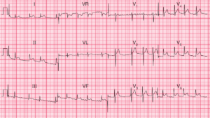
ECG Interpretation
- Sinus rhythm, rate 64/min
- Short PR interval at 100 ms
- Normal axis
- Normal QRS complex duration
- Slurred upstroke to QRS complexes (delta wave)
- Dominant R wave in lead V1
- Normal ST segments and T waves
Clinical Interpretation
This ECG shows the Wolff–Parkinson–White (WPW) syndrome type A, which is characterized by a dominant R wave in lead V1.
What to do ?
The catch here is that the dominant R wave in lead V1 may be mistakenly thought to be due to right ventricular hypertrophy. In a young woman who complains of breathlessness after a pregnancy, pulmonary embolism is obviously a possibility, and this might well cause ECG evidence of right ventricular hypertrophy – but in the presence of the WPW syndrome this would be very difficult to diagnose from the ECG. The only thing that might help the diagnosis would be the appearance of right axis deviation, which is not part of the WPW syndrome, and is not present here. So, look for another cause of breathlessness, such as anaemia.