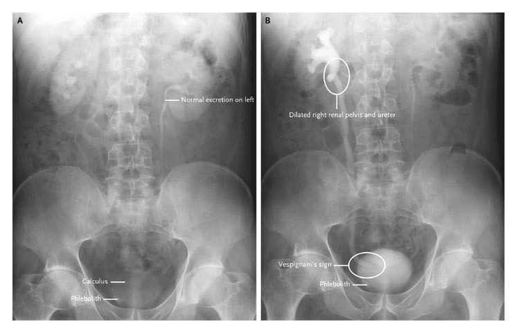
A 57-year-old man was referred to our hospital because of an acute onset of right flank pain. An intravenous urogram showed normal excretion from the left kidney, a calculus in the right pelvis, and a phlebolith in the right pelvis at 3 minutes after injection of the contrast material (Panel A).
Then showed delayed excretion of the contrast material by the right kidney, a dilated right renal pelvis and ureter to the bladder, and a filling defect around the ureteric meatus at 12 minutes after injection (Panel B). The findings are related to edema due to the passage of a calculus.
These findings are most suggestive of a calculus at the right uretero-vesical junction.
After treatment with a nonsteroidal antiinflammatory drug, the pain resolved and the patient excreted a calculus. He was then discharged from the hospital.
READ ALSO : Comprehensive Urolithiasis Guidelines

