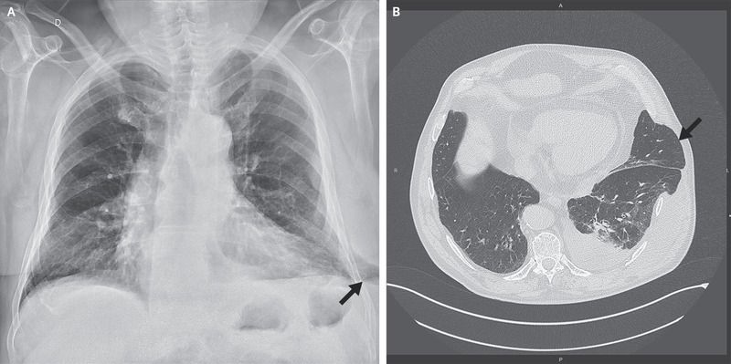
A 67-year-old man was admitted to the emergency department with a 10-day history of pain in the left side of the chest that had begun after an episode of severe coughing. He described persistent pain, without dyspnea, that was unrelieved by analgesic agents. He also noted swelling on the lower left side of the chest.
The patient had a history of smoking, hypertension, and chronic obstructive pulmonary disease that required treatment with continuous positive airway pressure. There was no history of chest trauma or surgery involving the chest.
Physical examination revealed thoracic asymmetry, with a soft, reducible mass corresponding to the region of swelling that was painful on palpation. The mass increased in size with inspiration and decreased in size with expiration.
A radiograph of the chest showed extension of lung parenchyma beyond the rib cage laterally at the base of the left lung (Panel A, arrow). Computed tomography of the chest revealed lung herniation through a left lower intercostal space laterally, as well as a moderate pleural effusion on the left side (Panel B, arrow). The patient underwent successful surgical repair of the hernia.

