This post is an answer to the ECG Case 294
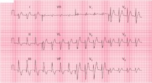
- Rate: 72 bpm
- Rhythm: Regular
- Axis: Right axis deviation
- Intervals:
- QRS – Prolonged (120ms)
- QT – Measured in lead aVL 440ms
- QT – Measured in leads V4-5 620-640ms
- Segments:
- ST Elevation in leads aVR, aVL, V1-3
- ST Depression in leads II, III, aVF, V4-6
- Additional:
- U waves best seen in precordial leads
- Associated apparent QT prolongation in precordial leads vs limb leads due to T-U fusion
- T wave inversion in leads II, III, aVF, V4-6
- LVH criteria
- V4 R >26mm
- Largest Precordial S + R wave >45mm
- R wave in aVF ~20mm
Interpretation
- Features of hypokalaemia
- Prominent U waves
- Apparent QT prolongation due to T-U fusion in precordial leads
- Cause of / or contributing to T wave inversion and ST depression
- T wave and ST segment changes could be due to LVH
- Potential for ACS
- Needs serial ECG’s
What happened next ?
Patient’s potassium was 2.1 mmol/L. Serial troponins were negative. Partial resolution of ST segment depression and resolution of U waves once K was corrected.
READ MORE:
SIMILAR CASES: