This post is an answer to the ECG Case 295
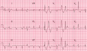
- Rate: 66 bpm
- Rhythm: Regular, Sinus Rhythm
- Axis: Normal (-33 deg)
- Intervals:
- PR – Normal (~160ms)
- QRS – Normal (100ms)
- QT – 380ms (QTc Bazette 380-400 ms)
- Segments:
- Subtle ST depression in leads V4-6
- Subtle ST elevation in aVL and V2
- Additional:
- T wave inversion in lead III
- Biphasic T wave lead aVF
- Prominent T waves in leads I, aVL, V2 (of equal or greater height than QRS)
Interpretation
Very suspicious ECG for ACS
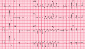
What happened next ?
The patient had serial ECG’s which showed dynamic T wave changes in the inferior leads and T wave amplitude antero-laterally. Serial troponins were positive. The patient underwent angiography which showed:
- LMCA: Minor irregularities
- LAD: Mid 99% single discrete lesion
- Ostial 1st Diagonal: 90% single discrete lesion
- CX: Irregularities
- RCA: Irregularities
A stent was inserted to the LAD lesion and the ostial lesion was treated with balloon angioplasty. Echo showed normal systolic and valvular function. The patient was commenced on dual anti-platelet therapy (DAPT), statin, ACE and beta-blocker therapy.
READ MORE: ECG Interpretation – All you need to know