This post is an answer to the ECG Case 307
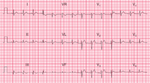
- Rate: 78 bpm
- Rhythm: Sinus arrhythmia
- Axis: Normal
- Intervals:
- PR – Normal (~180ms)
- QRS – Normal (80-100ms)
- QT – 380ms (QTc Bazette 435 ms)
- Segments:
- ST Elevation in leads I and aVL (~1mm)
- ST Depression in leads II, III, aVF, V4-5
- Additional: Prominent T wave in lead V2

Interpretation
High lateral STEMI
What happened next ?
Patient was taken for urgent angiography which showed:
- Left main: Irregularity
- LAD: Diffuse disease 60% mid
- D1: 90% Mid
- LCx: 90% Proximal 70% Mid – PCI to LCx
- OM3: 70% Ostial
- RCA: Dominant 70% Ostial long 40-50% Mid
Post angio echo showed normal LV function and size with apico-lateral hypokinesis. The patient made an uneventful post procedural recovery and will likely need further PCI to RCA and LAD.
READ MORE: ECG Interpretation: All you need to know
SIMILAR CASE: ECG Case 297