This post is an answer to the ECG Case 317
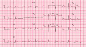
- Rate: 72 bpm
- Rhythm: Regular sinus rhythm
- Axis: Normal
- Intervals:
- PR – Normal (~160ms)
- QRS – Normal (100ms)
- QT – 360ms
- Segments:
- ST Elevation in leads aVR (1mm), aVL (1mm), V1 (1mm), V2
- ST Depression in leads II, III, aVF, V4-6
- Additional:
- Prominent U-wave in antero-septal leads
- T wave inversion infero-lateral leads
- Down-up morphology may be due to prominent U waves
Interpretation
- Acute OMI
- Patient with history suspicious of ACS
- ST / T changes indicative of OMI
What happened next ?
The patient was taken for urgent angiography which showed:
- Right dominant system
- LM: 50% distal
- LAD: 90% proximal
- Cx: 90% mid
- RCA: 99% distal RCA with 80% ostial – TIMI 3 flow and pain-free patient
- RCA: Supplying large PDA and 3 PLV branches
- LH Cath: Inferior akinesis with mild LV impairment
The patient was then transferred to tertiary centre for urgent CABG given severe multi-vessel disease.
SIMILAR CASES: