Interpretation
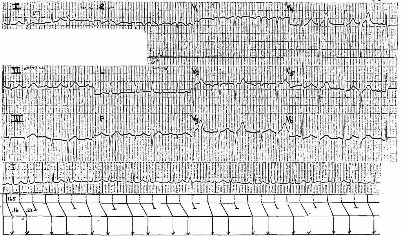
The patient has Atrial Tachycardia at 165/min with 3:2 AV Block Mobitz I Wenckebach.
The second of the conducted P waves is transmitted with Left Anterior Fascicular Block (LAFB) aberration.
Note that the first conducted QRS shows QS pattern in lead III and AVF consistent with Old Inferior MI.
Look what happend when LAFB occurs. Now there are small, but discrete R waves in lead III and AVF, and no R waves in V1, V2, V3. The LAFB has masked the evidence of Inferior MI and made it look like Anterior MI.
Explanation for these changes:
- Inferior MI redirects initial forces left and superior – away from the Infarct zone. With LAFB the initial forces become directed inferiorly. When these two abnormalities are combined, the evidence of prior Inferior MI disappears.
- Recall that a synonim for the left anterior fascicle is left superior fascicle. LAFB will result in initial forces that are redirected inferior-posterior. The normal position of the V1, V2, V3 electrodes may “see” the initial vector oriented away from the recording electrode
Read More About: ECG Interpretation


