ECG Interpretation
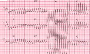
The lead II rhythm strip at the bottom of the record shows that the rhythm changed during the recording, so it is necessary to try to identify the normal complexes (if any) in each lead.
There are normal beats in the second and third complexes in leads I, II and III; in the first complex in leads VR, VL and VF; in the last complex in leads V1–V3; and in the first complex in leads V4–V6. The ECG shows:
- Sinus rhythm at about 77/min, with ventricular extrasystoles at the beginning and end of the record, and a six-beat run of a broad complex rhythm in the middle of the record
- The first complex of the run of broad complexes differs from the others, and is probably a fusion beat (a combination of a sinus beat and the ectopic rhythm)
- Normal axis when in sinus rhythm
- Normal QRS complexes in sinus rhythm
- Inverted T waves in lead III, but not VF
Clinical Interpretation
The run of broad complexes represents accelerated idioventricular rhythm. This is quite common following a myocardial infarction, but in a healthy subject it is probably of no significance. The T wave inversion in lead III is not important because the T wave is upright in lead VF.
What to do ?
If the individual has no symptoms and the physical examination is normal, no further action is needed. Accelerated idioventricular rhythm should not be treated.