ECG Interpretation
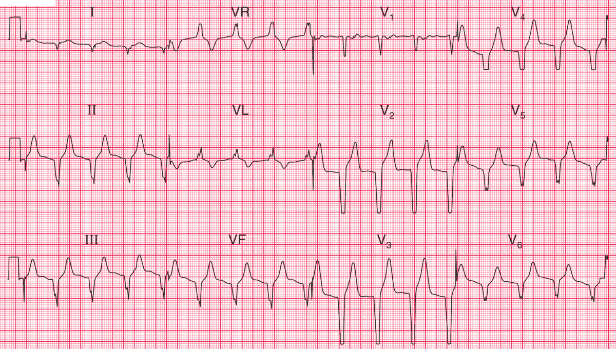
- Broad complex rhythm, rate 90/min
- No P waves
- Marked left axis deviation
- Positive QRS in AVR
- QRS complex duration 160 ms
- All chest leads show a downward QRS complex (concordance)
Clinical Interpretation
If the heart rate were fast there would be little difficulty in recognizing this as ventricular tachycardia, and this rhythm used to be called ‘slow VT’. It is, however, an accelerated idioventricular rhythm.
What to do ?
This rhythm is quite commonly seen in patients with an acute myocardial infarction, and indeed is not uncommon in ambulatory ECG records from normal people. It never causes problems, and it is important not to attempt to treat it: suppressing any ‘escape’ rhythm may lead to a dangerous bradycardia.


