This article is an answer to the ECG Case 176
The rhythm is regular at a rate of 72 bpm. There are P waves (*) before each QRS complex with a stable PR interval (0.20 sec). The P wave is positive in leads I, II, aVF, and V4-V6 and negative in lead aVR. The P-wave morphology is normal. This is a normal sinus rhythm.
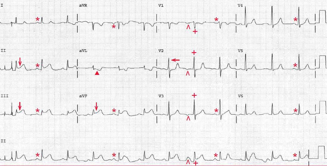
The eighth QRS complex is early (+) and the P wave before it (^) has a different morphology (ie, it is negative in leads V1-V3) compared with the other P waves. This is a premature atrial complex.
There is early transition with a tall R wave (←) in lead V2. This is termed counterclockwise rotation, determined by imagining the heart as viewed from under the diaphragm. When there is counterclockwise electrical rotation, left ventricular forces are seen earlier in the precordial or chest leads, accounting for early transition with a tall R wave in lead V2.
There is ST-segment elevation (↓) in leads II, III, and aVF, and the ST segments have a normal convex morphology. This is an acute inferior wall ST-segment elevation myocardial infarction (STEMI). ST-segment depression (▲) can be seen in lead aVL, which represents a reciprocal change.