This post is an answer to the ECG Case 201
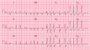
- Rate: 60 bpm
- Rhythm:
- Sinus Arrhythmia
- Axis:
- Normal (50 deg)
- Intervals:
- PR – Prolonged (~220ms)
- QRS – Normal (100ms in lead II, prolonged in lead V2)
- Apparent QT – 680ms (QTc Bazette ~ 710 ms)
- Segments:
- ST Depression in Leads I, II, V2-6
- ST Elevation in aVR
- Additional:
- Ventricular Ectopic
- Prominent U waves
- T-U Fusion
- Best visualised in leads II, III, aVF, V2-6
- Initial T wave is inverted and merges with large U wave
- Results in apparent QT prolongation due to fusion
- Best considered QU prolongation

Interpretation
Multiple ECG features consistent with hypokalaemia +/- hypomagnesaemia. This patient had a K+ of 1.6 mmol/L confirmed by a VBG.
READ MORE: Hypokalemia ECG Changes [With Examples]
SIMILAR CASES: