This post is an answer to the ECG Case 227
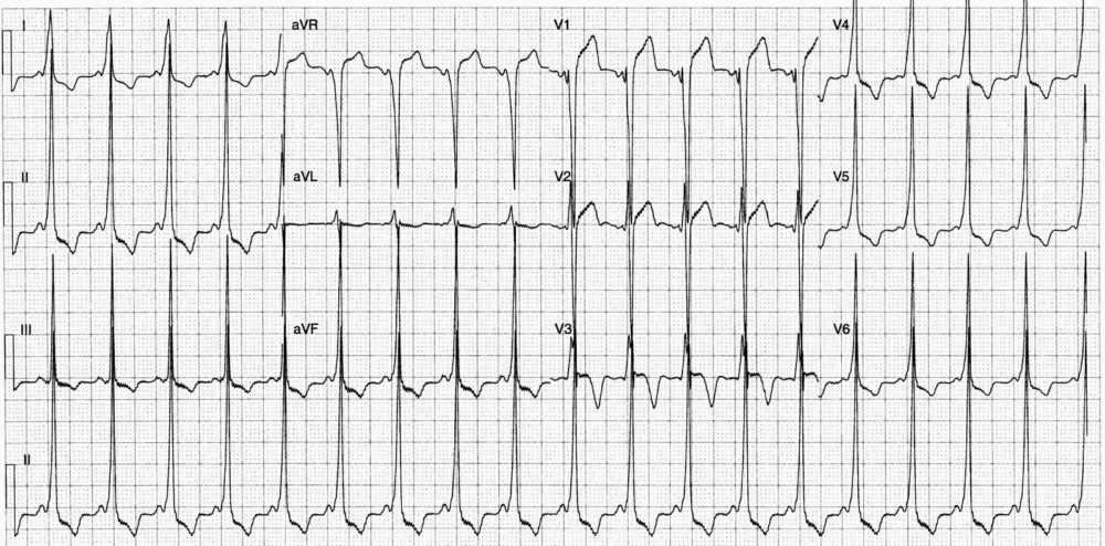
- Rate: 110-115 bpm
- Rhythm:
- Regular
- Sinus rhythm
- Axis: Normal
- Intervals:
- PR – Short (80ms)
- QRS – Prolonged (120ms)
- QT – 340ms (QTc Bazette 460 ms)
- Segments:
- ST Elevation leads in aVR, V1-2
- ST Depression in leads I, II, III, aVF, V4-6
- Additional:
- Delta waves best seen inferolaterally
- T wave inversion in leads I, II, III, aVF, V3-6
- ‘Pseudo’ left ventriclar hypertrophy
- Prominent R waves in leads I, II, III, aVF, V4-6
- Deep S waves in leads aVR, aVL, V1-2
Interpretation
- Wolff-Parkinson-White
- Right anteroseptal pathway – using Arruda algorithm
- Voltage & ST/T changes secondary to pre-excitation
- Patient requires referral for an EP study.
SIMILAR CASES:


