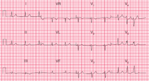
This post is an answer to ECG Case 188
- Rate: 70
- Rhythm:
- Sinus Rhythm
- Sinus Arrhythmia
- Axis:
- Normal (~70 deg)
- Intervals:
- PR – Short (~80-100ms)
- QRS – Prolonged (~120-160ms)
- QT – 400-440ms (QTc Bazette ~ 445-490 ms)
- Segments:
- ST Depression in V1-5
- Additional:
- T Inversion in V1-3, aVR
- Biphasic T wave in V4
- Slurring upstroke QRS
- Dominant R wave in V1 (R/S Ratio >1)
Interpretation
Short PR interval and Slurring QRS Upstroke (Delta wave) is consistent with Wolff-Parkinson-White (WPW).
Note changes similar to Right Ventricular Hypertrophy with strain – Dominant R wave in V1, R/S ratio > 1 in V1, ST depression & T inversion in anterior leads. These changes are seen in WPW due to pre-excitation and are not due to actual hypertrophy.
READ MORE:
SIMILAR CASES: