This post is an answer to the ECG Case 253
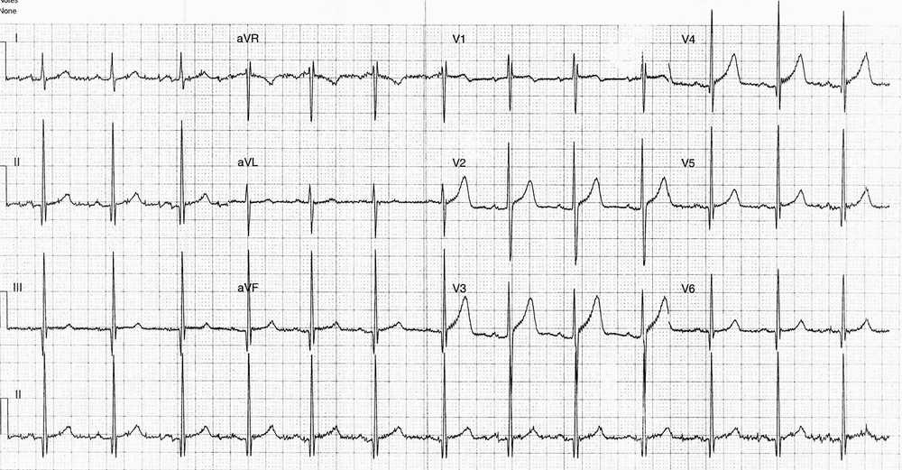
- Rate: 78 bpm
- Rhythm: Regular Sinus rhythm
- Axis: Normal
- Intervals:
- PR – Normal (~180ms)
- QRS – Normal (100ms)
- QT – 360ms (QTc Bazette 410 ms)
- Segments:
- Concave ST elevation in leads V2-4
- No ST depression
- Additional:
- Voltage criteria for LVH
- R wave V5 + S wave V1 ~35mm
- R wave aVF >20mm
- Narrow deep Q waves leads II, III, aVF, V4-6
- Partial RBBB pattern – rSr’ in lead V1
- Voltage criteria for LVH
Interpretation
Given the history of exertional syncope plus LVH with infero-lateral deep Q waves the major concern would be hypertrophic cardiomyopathy.
What happened next ?
The ECG changes were appreciated and the patient had a cardiology review and urgent echo. His echo was entirely normal and he was discharged with out-patient cardiology follow-up.
READ ALSO: Left Ventricular Hypertrophy (LVH): How to Recognize it on ECG [With Examples]


![Read more about the article Left Ventricular Hypertrophy (LVH): How to Recognize it on ECG [With Examples]](https://manualofmedicine.com/wp-content/uploads/2025/12/Summary-of-the-various-diagnostic-ECG-criteria-for-left-ventricular-hypertrophy-LVH.png)
