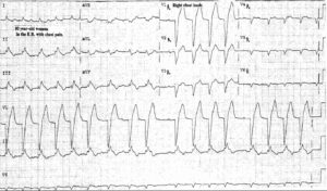
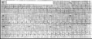
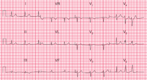
This post is an answer to the ECG Case 271
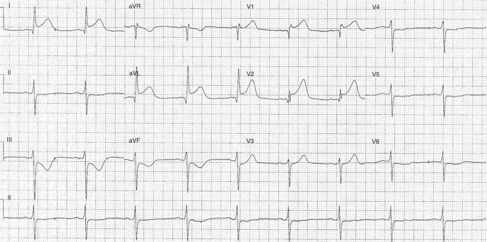
- Rate: 51 bpm
- Rhythm: Regular
- Axis: LAD
- Intervals:
- PR – Normal (180ms)
- QRS – Normal (100ms)
- Segments:
- ST Elevation in leads I (2mm), aVL (3mm), V1, (1.5mm),V2 (2mm)
- ST Depression in leads II, III, aVF, V4-6
- Additional:
- T wave inversion in II, III, aVF, V5-6
What happened next ?
The patient for taken for urgent angiogram which showed:
- 100 % Occlusion of the 1st diagonal branch –> Stented
- 40% stenosis mid LAD
- 50% stenosis distal LAD
- 70% stenosis distal CX
- 100%stenosis OM4
Echo showed apical anterior and anterolateral akinesis with normal systolic function. The patient made an uneventful recovery and was discharge after 2 days.