This post is an answer to the ECG Case 288
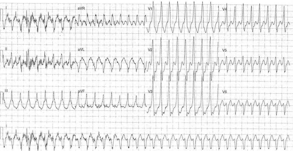
- Rate: ~210 bpm
- Rhythm: Regular
- Axis: RAD
- Intervals: QRS – Prolonged (120-160 ms)
- Additional:
- QRS Alternans – best seen in leads III & aVF
- Retrograde P waves buried in terminal QRS – best seen in V3
- Lack of concordance
- No fusion or capture beats
- Marked baseline artifact in leads I, II, aVR
Interpretation
Wide complex tachycardia (WCT). General differentials for all WCT include:
- VT
- SVT with aberrancy (rate ralted BBB, pre-exisiting BBB)
- SVT with pre-excitation
- Paced rhythm
Given the patient’s age the likeliest diagnosis is one of SVT.
The patient was treated with a valsalva maneuver which resulted in cardioversion.
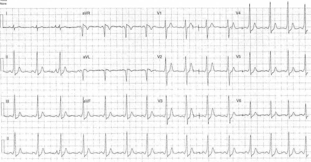
- Rate: 90 bpm
- Rhythm: Regular sinus rhythm
- Axis: Inferior axis
- Intervals:
- PR – Short (120ms)
- QRS – Prolonged (110-120 ms)
- QT – 320ms
- Segments:
- ST Elevation in lead aVR and aVL
- ST Depression in leads II, III, aVF, V1-6
- Additional:
- Delta waves – best seen in leads II, III, aVF, V1-6
- Deep Q wave in lead aVL – ‘pseudo-infarction’ pattern due to pre-excitation
- Artifact mimicking pacing spikes in leads V1-3
Interpretation
WPW (Left lateral / anterolateral accessory pathway (Arruda Algorithm)) and an episode of antidromic AVRT
READ MORE: Never Mistake Ventricular Tachycardia for Supraventricular Tachycardia with Aberrant Conduction
SIMILAR CASE: ECG Case 88 – Antidromic AVRT with WPW


