This post is an answer to the ECG Case 298
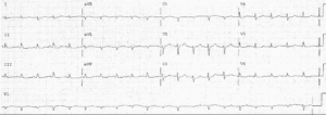
- Rate: 72 bpm
- Rhythm: Regular
- Axis: Normal
- Intervals:
- PR – Normal (140ms)
- QRS – Normal (100ms)
- Segments:
- Cove-shaped ST elevation in lead V1 (<1mm) and V2 (3mm)
- Obliquely straight ST elevation V3 (4mm)
- Possible ST depression leads III, aVF – difficult to see due to baseline artifact
- Additional:
- P waves difficult to see due to baseline artifact – best seen in V2-3
- T wave inversion in leads aVR, aVL, V1-2
Interpretation
- Brugada Syndrome
- Type 1 Pattern
- Given the history of central chest pain the presence of additional ACS must be considered especially given the flat ST elevation in V3 plus subtle inferior ST depression
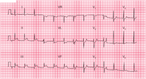
This is a challenging ECG, serial ECG’s and comparison with old ECG’s are essential. This patient had known Brugada and thankfully brought his old ECG from 10 years ago with him which showed all changes to be longstanding.
What happened next ?
Interestingly the patient had been lost to cardiology follow-up and had no AICD inserted for his Type 1 Brugada. Given the typical nature of the pain the patient had a coronary angiogram which revealed no evidence of coronary artery disease and he is awaiting an AICD insertion.
READ ALSO: Pearls in Syncope ECG Interpretation
SIMILAR CASES: