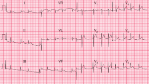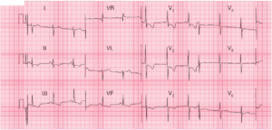
This post is an answer to the ECG Case 341
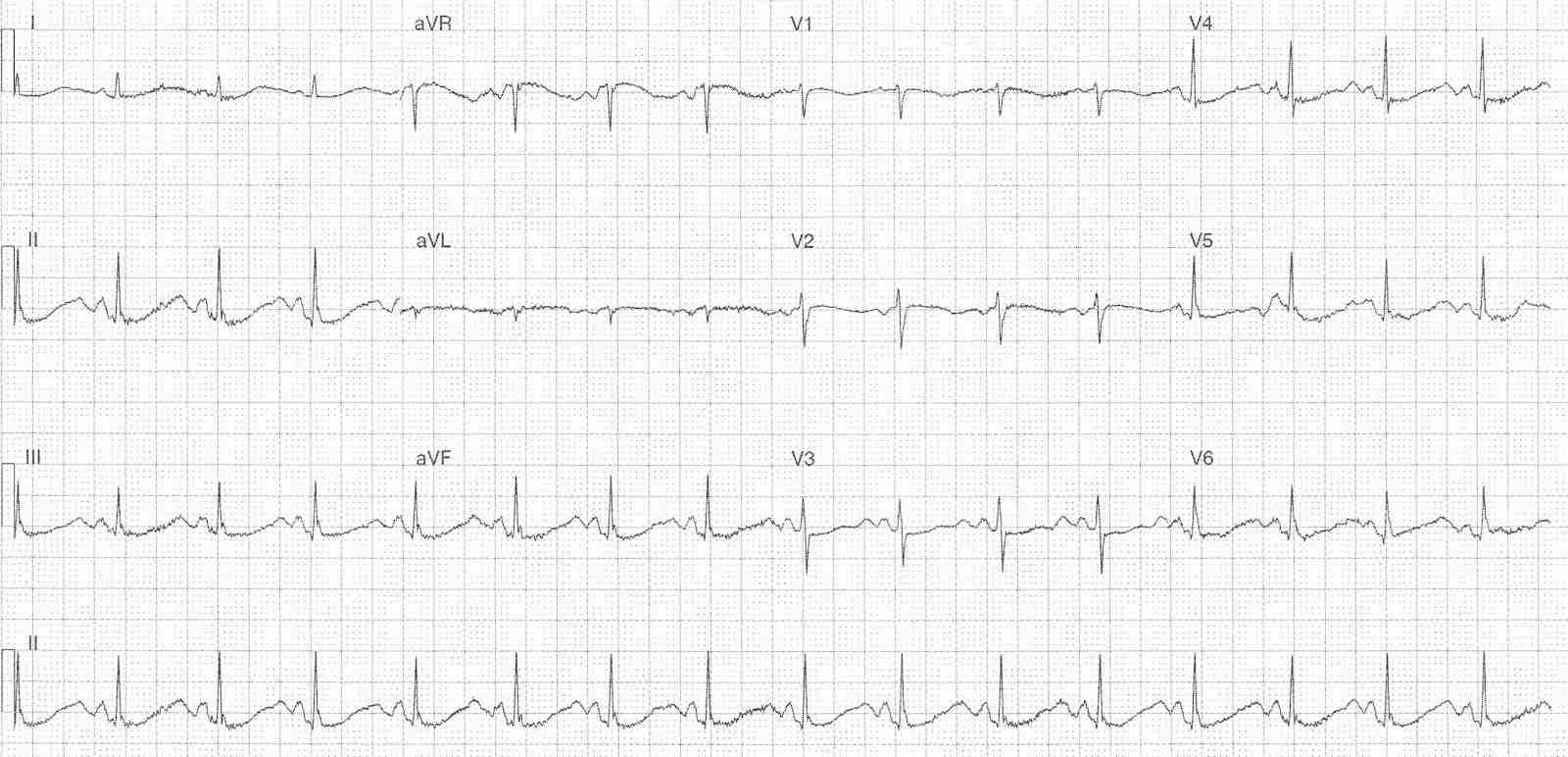
- Rate: 96 bpm
- Rhythm: regular sinus rhythm
- Axis: normal
- Intervals:
- PR – Normal (~180ms)
- QRS – Normal (80ms)
- QT – 500-520ms (QTc Bazette 630-660 ms) measured in lead II
- Additional:
- ST depression in leads II, III, aVF, V4-6
- Prominent U waves in leads V3-5 just before the p wave
- T-U fusion in all other leads
Interpretation
- Marked QT Prolongation
- Features supportive of hypokalemia / hypomagnesemia
- U waves T-U fusion
- Variable QT measurement lead II vs lead V3 (end of T wave more easily identifiable)
- Potential contribution from sotalol – known QTc prolonging agent
What happened next ?
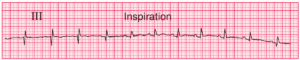
Shortly after this ECG was performed the patient became unresponsive with the following ECG rhythm strip.
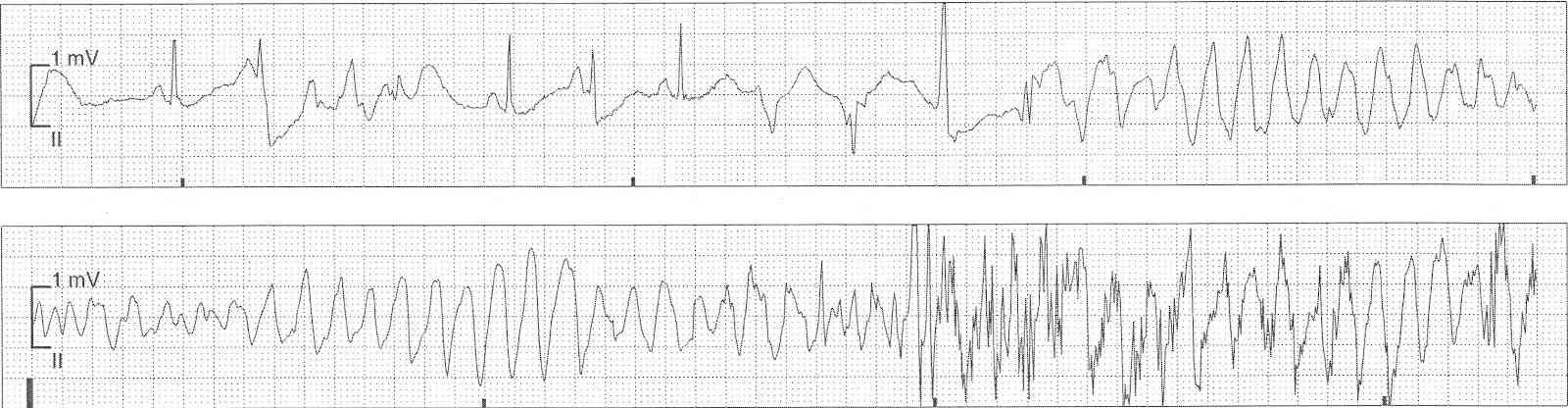
The rhythm strip shows sinus rhythm with several PVC’s with resultant R-on-T phenomenon and degeneration into polymorphic VT. This episode was brief and self-terminated. Bloods revealed several metabolic alkalosis, hypokalaemia and hypomagnesaemia.
The patient was admitted to a critical care area for monitoring and correction of electrolyte and acid-base disturbance. In addition, her sotalol was ceased due to its associated risk of QTc prolongation and she was placed on metoprolol.
READ MORE: Hypokalemia ECG Changes [With Examples]
SIMILAR CASES: