This post it an answer to the Case – Chest Pain in a Patient with History of Bone Marrow Transplant
Findings
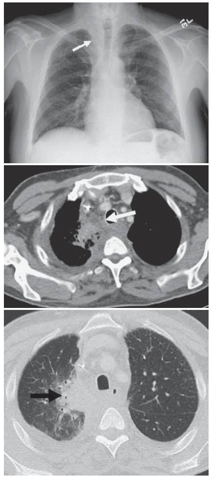
- The chest radiograph demonstrates a right paratracheal soft tissue density with mass effect on the trachea (white arrow). This mass has developed in a 2-week interval.
- CT images demonstrate a right upper lobe consolidation (white arrow) . There is loss of the normal fat planes within the mediastinum accompanied by an increase in attenuation of the mediastinal fat at the level of the consolidation (black arrow).
Differential Diagnosis
Rapid increase in mediastinal size can be seen in the setting of hematoma and mediastinitis. The clinical history can be very helpful in making the distinction. The absence of trauma and recent line placement would favor the latter. Rarely, aggressive leukemia or lymphoma can present with change in the mediastinal size in less than 1 month.
Teaching Points
- Acute mediastinitis usually results from perforation of the esophagus or cardiac surgery, but may also result from extension of pneumonia, retropharyngeal infection, or sternoclavicular septic arthritis.
- Acute necrotizing mediastinitis has a very high mortality, with reported rates of 30% to 50% .
- CT is helpful in evaluating the presence of fluid collections that may need to be drained.
- Occasionally on CT pneumomediastinum may be seen. Mediastinal fat stranding and enlargement are almost always present.
- The postoperative mediastinum can be very difficult to distinguish from acute mediastinitis. Fluid and gas collections in the noninfected, postoperative mediastinum, however, should begin to resolve by 3 weeks.
- Particular attention needs to be paid for the evaluation of an esophageal perforation, as this requires urgent surgical intervention and carries a high mortality even if treated.
Management
Treatment is based on correcting the underlying etiology (perforated esophagus, septic arthritis, cellulitis) but may also consist of percutaneous drainage if any abscesses are present. Antibiotics are always instituted.
Further Reading
- Athanassiadi KA. Infections of the mediastinum. Thorac Surg Clin. 2009 Feb;19(1):37-45, vi. doi: 10.1016/j.thorsurg.2008.09.012. PMID: 19288819.
- Exarhos DN, Malagari K, Tsatalou EG, Benakis SV, Peppas C, Kotanidou A, Chondros D, Roussos C. Acute mediastinitis: spectrum of computed tomography findings. Eur Radiol. 2005 Aug;15(8):1569-74. doi: 10.1007/s00330-004-2538-3. Epub 2004 Dec 31. PMID: 15627194.