This post is an answer to the Case – Shoulder Pain in a 49-year-old Woma
Findings
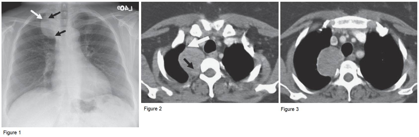
- The radiograph demonstrates a right paratracheal mass extending above the clavicle (white arrow in Fig. 1). Splaying of the posterior right fourth and fifth ribs is also seen.
- Black arrows in Figure 1 show the interface of the mass with the lung (mediastinal border), which creates an obtuse angle, suggesting a mediastinal lesion.
- CT ( Figs. 2 and 3 ) confirms the mass to be posterior mediastinal. The mass subtly enhances (black arrow in Fig. 2). White arrow in Figure 2 shows that the interface with the lung creates an obtuse angle.
Differential Diagnosis
Differential diagnosis is based on a posterior mediastinal mass. In an adult, a nerve sheath tumor would lead the differential diagnosis. Other causes of posterior mediastinal masses, including extramedullary hematopoiesis, lateral meningocele, and vertebral osteomyelitis/diskitis are not likely.
Teaching Points
- The cervicothoracic sign refers to the superior extension of the mass above the clavicles. Because the anterior lung stops at the level of the clavicles, a mass demonstrating this sign must be posterior in the thorax or arising from the neck.
- Posterior mediastinal lesions are considered to be neurogenic until proven otherwise. In an adult, they tend to be of nerve sheath origin (neurofibroma, schwannoma, or neurolemmoma). In children and young adults, they may be of sympathetic ganglia origin (ganglioneuroma, neuroblastoma, ganglioneuroblastoma).
- Nerve sheath tumors tend to be one or two rib interspaces in z-axis, while sympathetic ganglia tumors are longer.
- Schwannomas and neurofibromas are well circumscribed and round. Because of myelin and occasional cystic regions, they tend to be lower than muscle in CT attenuation. On MR, they tend to be higher than muscle on T2 and often enhance with a targetoid appearance.
- Neural foraminal enlargement may not be seen with nerve sheath tumors, but osseous remodeling is common.
- Schwannomas are usually incidental, but one third of patients with neurofibromas will have neurofibromatosis.
Management
These may be resected because of local symptoms. The risk of malignant transformation is low in isolated schwannomas.
Further Reading
Whitten C, Khan S, Munneke G, et al. Diagnostic approach to mediastinal abnormalities. Radiographics. 2007; 27:657-671.