
This article is an answer to the Case – 30 Years Old Man Involved in Motor Vehicle Accident
CT scan findings
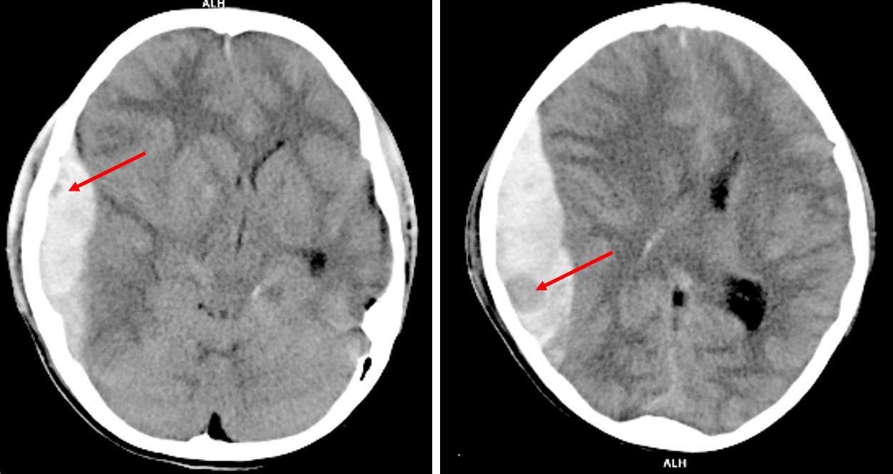
- There is subdural hemorrhage at right fronto-temporo-parietal region
- Areas of hypodensity seen (red arrows) within the hemorrhage representing Swirl Sign
- There is midline shift to the left side
- Compression of ipsilateral lateral ventricle
Swirl Sign
- Swirl sign is seen in non-contrast CT brain in acute extradural or subdural hemorrhage.
- It represents unclotted blood which is hypodense compared to surrounding clotted blood which is hyperdense (50-70HU).
- It has been verified as active bleeding site at the time of surgery
- Identification of patients with a potential risk for hematoma expansion in acute intracranial hemorrhage is crucial for planning of the treatment and prediction of the functional outcome.
- Swirl sign is an indicator of possible hematoma expansion and associated with higher mortality compared to patient without this sign.