This post is an answer to the Case – Smoker with Haemoptysis and Weight Loss
Chest X-Ray Interpretation
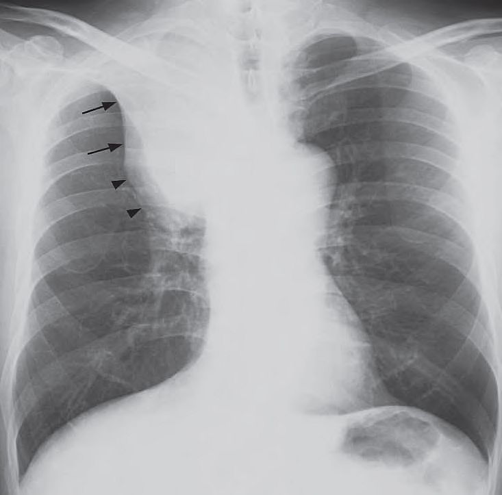
- Opacity with a sharp well-demarcated lateral border (arrows) in right upper zone with lack of air within the abnormality.
- Focal convex bulge at the apex of the abnormality.
- Hyperinflation of the right lower lobe.
- Elevated right hemidiaphragm.

Diagnosis
The ‘Golden S sign’ – collapse of the right upper lobe with a well demarcated lateral border formed by the elevated horizontal fissure (arrows), and a focal convex bulge at the apex due to the centrally located bronchogenic carcinoma (arrowheads).
READ MORE: Cough and Hemoptysis – Differential Diagnosis, Examination and Investigations

