This post is an answer to the Case – 28-year-old Woman with Cardiac Disease and Dyspnea
Findings
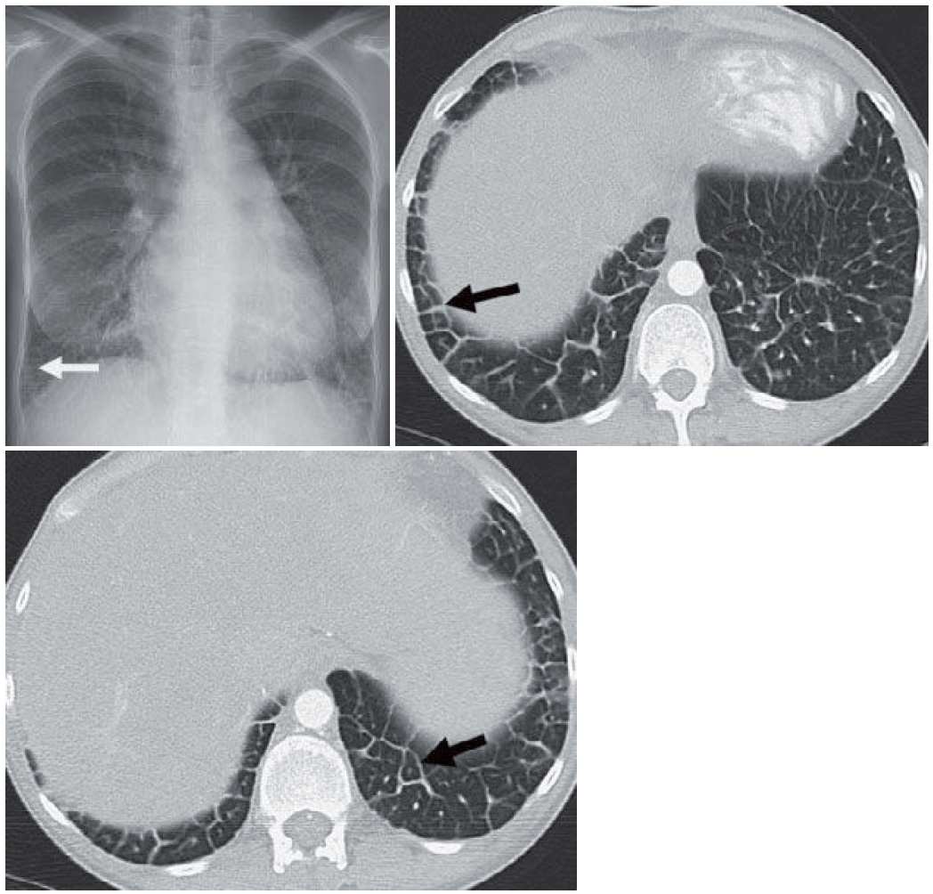
- Chest radiograph shows bilateral septal lines (Kerley B lines) (arrow).
- CT confirmed smooth interlobular septal line thickening (arrow). The smooth septal lines outline the secondary pulmonary lobules (arrow).
Differential Diagnosis
The differential diagnosis for smooth septal line thickening is based on acuity. Acutely, smooth septal lines are usually seen in hydrostatic or cardiogenic edema. They can also be seen in hemorrhage (which usually has accompanying ground glass) or viral pneumonias (which usually have ground-glass nodules). More chronic smooth septal line thickening can be seen with lymphangitic carcinomatosis and, very rarely, amyloid or lymphoma. Given the history of cardiac disease, cardiogenic edema should be favored.
Teaching Points
- Hydrostatic pulmonary edema refers to the presence of interstitial fluid within the lungs, usually from left heart failure (cardiogenic pulmonary edema). Other causes include isolated pulmonary venous obstruction, fluid overload, and hypoalbuminemia.
- Pleural effusions and cardiomegaly may be seen in cardiogenic edema.
- Radiographic findings of cardiogenic edema include interlobular septal line thickening, peribronchial cuffing, ground-glass opacities, and frank consolidation.
- Cardiogenic pulmonary edema does not follow a programmed course from interstitial edema to consolidation. Patients can present acutely with either interstitial or airspace disease.
- Peripheral 1 to 2-cm interlobular septal lines are sometimes referred to as Kerley B lines. Kerley A lines are central interlobular septal lines that are longer.
- Cardiogenic edema is usually bilateral. Unilateral edema should prompt inspection of central structures to look for a mass that may be compressing the pulmonary vein. Most of the time, unilateral pulmonary edema develops because of patient positioning.
- Cardiogenic edema should clear rapidly as the patient improves clinically. When it does not change, alternative diagnoses should be considered, such as lymphangitic carcinomatosis or, in the setting of ground glass, diff use alveolar damage.
Management
Cardiogenic edema is usually treated with supportive therapy and diuretics. Once the patient is through the acute situation, attempts are made to maximize cardiac function.
Further Reading
Webb WR. Thin-section CT of the secondary pulmonary lobule: anatomy and the image–the 2004 Fleischner lecture. Radiology. 2006 May;239(2):322-38. doi: 10.1148/radiol.2392041968. Epub 2006 Mar 16. PMID: 16543587.