This article is an answer to the case – Primigravid Woman with Proximal Muscle Weakness, Striae, and Facial Plethora
An abdominal ultrasound examination, performed as part of the evaluation for Cushing’s syndrome, revealed a right adrenal mass that was 7 cm in length in its largest dimension.
The level of free cortisol in the urine over 24 hours was 3058 nmol (reference value, <160). The patient’s hypertension was managed with labetalol and nifedipine; however, HELLP syndrome (characterized by hemolysis, elevated liver enzymes, and low platelet count) developed at 34 weeks of gestation. A healthy infant weighing 2890 g was delivered.
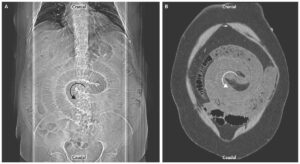
A computed tomographic scan obtained without contrast material showed an adrenal mass measuring 6.8 cm in its largest dimension (image) with a density of 22 Hounsfield units, a finding suggestive of adrenal carcinoma.
Adrenalectomy was performed 1 month after delivery, and histopathological analysis confirmed the presence of high-grade adrenocortical carcinoma.
An evaluation showed no evidence of metastatic disease, and treatment with adjuvant mitotane was initiated.