This article is an answer to the Case – Ptosis and Anisocoria after Traumatic Head Injury
An examination of his pupils at his arrival showed ptosis (Panel A) and anisocoria (Panel B), which aroused concern that there was uncal herniation.
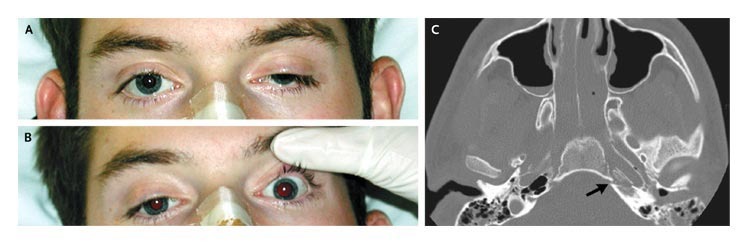
A high-resolution computed tomographic scan of his head did not show uncal herniation but, rather, revealed a complex fracture of the sella turcica (Panel C, arrow) and of the skull base (not shown), with impingement of the bony nerve canals and a C1 fracture.
He was discharged to his home 16 days after the injury with a hard cervical collar. Subsequently, he has done well but has had occasional vertigo and tinnitus. His ocular defects have resolved.
References
- Hae Won Noh, Jae Young Song, Jong Hyun Kim, Jang Hun Kim. (2017) Bilateral Oculomotor Nerve Palsy after Head Trauma: A Case Report. Journal of Trauma and Injury 30:2, 66-69.
- Ernest E. Wang. 2013. Cranial Nerve Disorders. Emergency Medicine, 818-829.e1.
- (2006) Traumatic Cranial Nerve Palsy. N Engl J Med 354:10, 1096-1096.