This post is an answer to the Case – Lifelong Neck Swellings
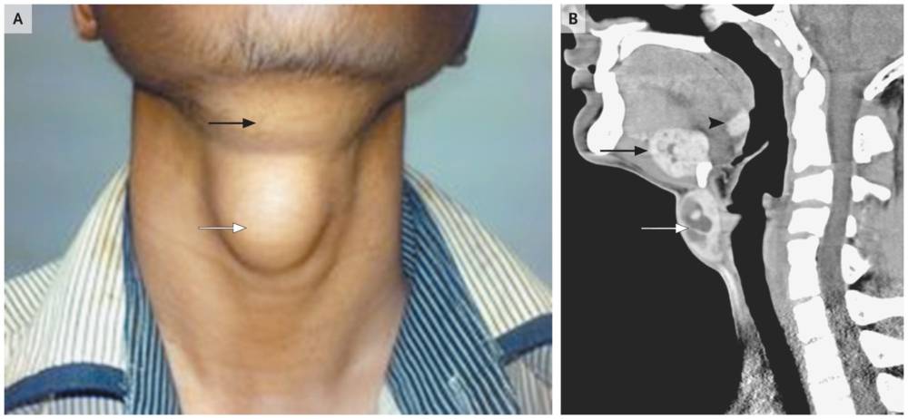
A 15-year-old boy presented with lifelong neck swellings that had been gradually increasing in size. He was otherwise asymptomatic. Physical examination was notable for a larger nodule in the infrahyoid area (Panel A, white arrow) and a smaller nodule in the suprahyoid area (Panel A, black arrow).
Laboratory tests revealed evidence of subclinical hypothyroidism, with elevated thyrotropin levels (12.6 μIU per milliliter; normal range, 2 to 11), accompanied by normal levels of triiodothyronine (92 ng per deciliter [1.4 nmol per liter]) and thyroxine (6.1 μg per deciliter [78.5 nmol per liter]).
After the administration of contrast material, computed tomographic scans of the neck showed homogeneously enhancing hyperdense lesions with nonenhancing cystic foci in the floor of the mouth (Panel B, black arrow) and between the hyoid bone and the thyroid cartilage (Panel B, white arrow). A third, smaller lesion with a similar pattern of enhancement was seen in the region of the foramen cecum (arrowhead).
The lesions in the three locations were clinically diagnosed as ectopic thyroid tissue. There was no normal thyroid gland in the customary location.
The parents of the patient decided against any intervention or further laboratory and radiographic evaluation. More than 1 year after presentation, the patient’s lesions continue to slowly increase in size. He remains otherwise asymptomatic.