This article is an answer to the Case – 23-year-old Man Involved in a Motor Vehicle Accident
The physical examination revealed multiple orthopedic injuries in addition to trauma to the right chest, pelvis, and head. He underwent prompt intubation and sedation.
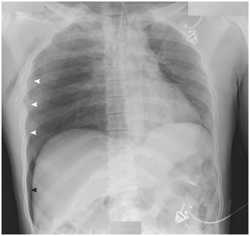
Chest radiography that was performed with the patient in the supine position showed a deep sulcus sign (black arrowhead), which was highly suggestive of a pneumothorax. On closer examination, a pneumothorax (white arrowheads) with associated rib fractures and subcutaneous emphysema were clearly evident.
The deep sulcus sign, which is characterized by a deep, lucent, ipsilateral costophrenic angle on supine chest radiography, is an indirect sign of a pneumothorax.
Intrapleural air distributes in a nondependent manner; thus, on upright radiographs, air collects in the apical lateral regions, and on supine radiographs intrapleural air collects initially in the anterior medial region and then in the lateral and caudal regions. The deep sulcus sign results from intrapleural air tracking in a laterocaudal manner.
A chest tube was placed, and a repeat chest radiograph revealed complete reexpansion of the lung. After a lengthy hospitalization and multiple orthopedic surgeries, the patient was discharged in stable condition.


