
A posteroanterior chest radiograph (Panel A) demonstrated a right-sided pneumothorax. His symptoms improved immediately on placement of a chest tube.
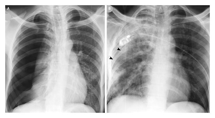
Two hours later, he again became breathless, and examination revealed extensive right-sided chest crackles. Chest radiography was repeated and showed a fully expanded right lung (Panel B), albeit with features of pulmonary edema. The arrowheads in Panel B show the position of the chest tube.
The patient’s condition improved after continuous positive airway pressure was delivered through a face mask overnight. The chest tube was removed after three days.
At follow-up six weeks later, the patient was asymptomatic and well. The results of further investigations were consistent with the presence of mild chronic obstructive pulmonary disease.