This article is an answer to the Case – A 4-year-old Girl with Watery Diarrhea, Progressive Abdominal Pain and Distention
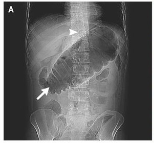
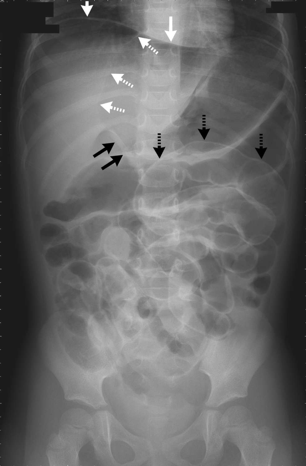
The laboratory evaluation revealed a white-cell count of 5000 per cubic millimeter, with 63% segmented neutrophils. The plain-film radiograph of the abdomen showed several signs of free intraperitoneal gas.
These included air accumulation in the right upper quadrant (the subphrenic area and ventral surface of the liver) (solid white arrows); the falciform-ligament sign, visible as a longitudinal linear density on the ventral surface of the liver (dashed white arrows); the ligamentum teres sign, visible as a linear density running along the inferior edge of the falciform ligament (solid black arrows); and Rigler’s sign, the visualization of air on both sides of the bowel wall (dashed black arrows). All these signs indicated pneumoperitoneum.
An explorative laparotomy revealed a cecal perforation caused by bowel-wall inflammation due to enteritis, as well as severe bowel dilatation. After repair of the perforation, the patient was discharged in good health 1 month later.
Similar Case: A 56-year-old man with Diarrhea and Epigastric Pain