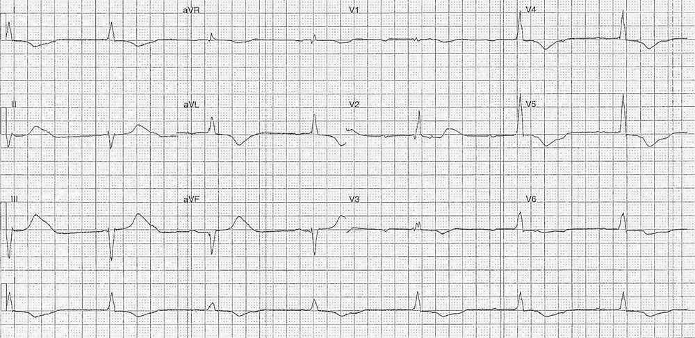
This post is an answer to the ECG Case 200
- Rate: 42/min
- Rhythm:
- Regular ventricular complexes
- Irregular atrial activity
- Complexes 3 & 4 sinus
- Axis:
- LAD (-50 deg)
- Intervals:
- PR – Normal (~180-200ms) for 3rd & 4th Complexes only
- QRS – Prolonged (~120ms)
- QT – 720ms (QTc Bazette ~ 600 ms)
- Segments:
- Slight down sloping ST Depression in V5-6
- Additional:
- T wave inversion in I, aVL, V1, V3-6
- QRS Morphology RBBB Pattern
- Differing QRS Morphology between complexes 1-2,5-7 and complexes 3-4
- Difficult to map atrial activity given relative low voltage p wave
- R wave progression abnormal across precordial leads
- Possible V2 misplacement – terminal deflection QRS larger than V1&2 with positive T wave
Interpretation
- Intermittent trifascicular block, likely complete
- Bifascicular block with variable sinus capture & high grade AV block
READ MORE about Conduction Blocks at the AV Node (AV Blocks) [With Examples]
SIMILAR CASES:


