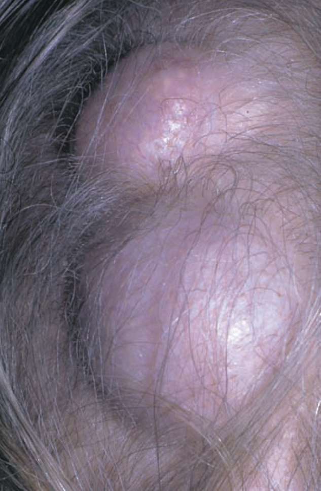
This post is an answer to the Case – Lumps on the Scalp
This has all the features of sebaceous cysts of the scalp – a very common lesion.
Describe the physical signs of a cystic swelling
- Fluctuation: The tip of the index finger of one hand is placed at the side of the lump, which is pressed upon by the tip of the opposite index finger – the ‘watching’ finger is felt to elevate. The test is repeated with the fingers moved at right angles to their previous position; again fluctuation is elicited.
- Transillumination: In addition, the lump can be shown to transilluminate when a bright torch is help against it, but this must usually be done in a darkened room. Transillumination is especially well seen in the examination of a ganglion, hydrocele or cyst of the epididymis.
Why bother to do the test for fluctuation in two directions at right angles to each other?
Try the experiment on yourself: ‘fluctuate’ transversally across the quadriceps muscle of your thigh – you can even do this experiment through your trousers or skirt – your watching finger will be elevated as you push the muscle mass transversally. Now try again with the fingers now at right angles, placed one above the other – now no movement is detected. You have not got cystic degeneration of your quadriceps!
Are there any other lumps that may occur on the scalp and which would therefore be a differential diagnosis to sebaceous cyst?
- An ivory osteoma of the skull vault is uncommon. It will not fluctuate, nor will it be moveable.
- Secondary deposits in the skull will give similar signs to an osteoma and there might well be clinical evidence of a primary tumour – breast in the female, prostate in the male and bronchus in both sexes are the headlines. Invasion of the skull by an underlying meningioma is a rarity.
Where else are sebaceous cysts found and where do they never occur?
Apart from the scalp, which is the commonest site, they are often found on the face, lobule of the ear, scrotum and vulva. They cannot occur on the sebaceous glandfree palms of the hands and soles of the feet.
What do these cysts look like when they are excised and cut open?
There is a white lining membrane (of squamous epithelium). The contents are white and cheesy, with an unpleasant smell.
What complications may they undergo?
Infection (the commonest), ulceration (‘Cock’s peculiar tumor’*), calcification, horn formation and malignant change (rare).
What treatment would you advise this woman to have?
Because of the common risk of infection, patients should always be advised to have their sebaceous cysts excised. This can be done quite simply under local anaesthetic. If acutely inflamed, surgical drainage is required, followed later by excision of the capsule wall to prevent further fl are-ups of infection. However, the inflammatory scar tissue now makes excision of the cyst a more difficult operation.