This post is an answer to the Case – Spinal Abnormality in a Newborn Child
What is this lesion called?
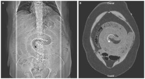
Spina bifida with meningocele – protrusion of the meninges through the vertebral defect without nervous tissue involvement.
How are the various types of this lesion classified?
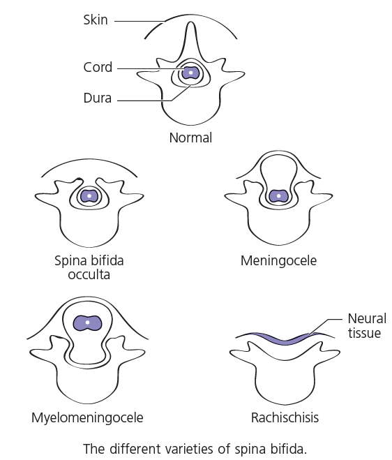
The following range of anomalies may be found, in ascending order of severity (image below):
- Spina bifida occulta: Failure of vertebral arch fusion only. This is symptom-less and is picked up incidentally on spinal X-ray. It may occur anywhere along the spinal column, but the great majority of defects involve L5 (6% of the population) or upper sacrum (11%). It may sometimes be associated with a tuft of hair, a lipoma or an overlying dimple in the skin.
- Meningocele (as in this case): Protrusion of the meninges without nervous tissue involvement.
- Myelomeningocele: Neural tissue (the spinal cord or roots) protrudes into and may adhere to the meningeal sac.
- Myelocele (or rachischisis): Failure of fusion of the neural tube, which results in an open spinal plate that occupies the defect as a red granular area weeping cerebrospinal fluid from its center. This condition is incompatible with survival.
What other congenital condition may typically coexist with this lesion?
Hydrocephalus occurs in 75% of cases of myelomeningocele (the Arnold–Chiari malformation). Hydrocephalus is very rare in an uncomplicated meningocele, such as in this child.
How should this lesion be treated?
The sac is excised and the skin defect repaired.