This post is an answer to the ECG Case 285
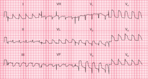
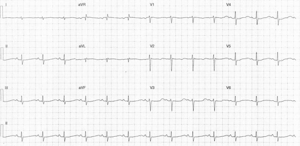
- Rate: 78 bpm
- Rhythm: Regular Sinus rhythm
- Axis: RAD (-33 deg)
- Intervals:
- PR – Normal (~170ms)
- QRS – Normal (80ms)
- Apparent QT – 560ms (QTc Bazette 640 ms)
- Due to T-U fusion
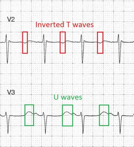
- See labelled image below
- Additional:
- Massive U waves
- Mimic T waves – see labelled image below
- Nearly merging into subsequent P waves
- T wave inversion in leads V1-2
- Massive U waves
Interpretation
ECG features consistent with hypokalaemia
What happened next ?
The patient had urgent electrolytes which showed:
- K 1.7 [3.4 – 5.5 mmol/L ]
- Cor Cal 2.45 [2.20 – 2.55 mmol/L]
- Mg 0.73 [0.70 – 1.20 mmol/L]
She was admitted to the HDU and a PICC line inserted due to high K replacement requirements. The patient made an uneventful recovery and was discharges once her potassium reached a consistently safe level.
READ MORE: Hypokalemia ECG Changes [With Examples]
SIMILAR CASES: